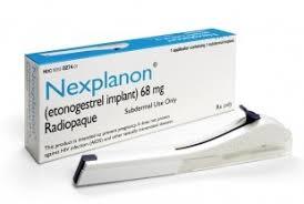
Nexplanon Dosage
Generic name: ETONOGESTREL 68mg
Dosage form: implant
Drug classes: Contraceptives, Progestogens
Medically reviewed by Drugs.com. Last updated on Dec 13, 2024.
The efficacy of NEXPLANON does not depend on daily, weekly, or monthly administration.
All healthcare professionals should receive instruction and training prior to performing insertion and/or removal of NEXPLANON.
A single NEXPLANON implant is inserted subdermally just under the skin at the inner side of the non-dominant upper arm. The insertion site is overlying the triceps muscle about 8-10 cm (3-4 inches) from the medial epicondyle of the humerus and 3-5 cm (1.25-2 inches) posterior to (below) the sulcus (groove) between the biceps and triceps muscles. This location is intended to avoid the large blood vessels and nerves lying within and surrounding the sulcus (see Figures 2a, 2b and 2c). Inserting an implant more deeply than subdermally (a deep insertion) may preclude palpation and localization, making removal difficult or impossible.
NEXPLANON must be inserted by the expiration date stated on the packaging. NEXPLANON is a long-acting (up to 3 years), reversible, hormonal contraceptive method. The implant must be removed by the end of the third year and may be replaced by a new implant at the time of removal, if continued contraceptive protection is desired.
Initiating Contraception with NEXPLANON
IMPORTANT: Rule out pregnancy before inserting the implant.
Timing of insertion depends on the woman's recent contraceptive history, as follows:
• No preceding hormonal contraceptive use in the past month
NEXPLANON should be inserted between Day 1 (first day of menstrual bleeding) and Day 5 of the menstrual cycle, even if the woman is still bleeding.
If inserted as recommended, back-up contraception is not necessary. If deviating from the recommended timing of insertion, the woman should be advised to use a barrier method until 7 days after insertion. If intercourse has already occurred, pregnancy should be excluded.
• Switching contraceptive method to NEXPLANON
Combination hormonal contraceptives:
NEXPLANON should preferably be inserted on the day after the last active tablet of the previous combined oral contraceptive or on the day of removal of the vaginal ring or transdermal patch. At the latest, NEXPLANON should be inserted on the day following the usual tablet-free, ring-free, patch-free or placebo tablet interval of the previous combined hormonal contraceptive.
If inserted as recommended, back-up contraception is not necessary. If deviating from the recommended timing of insertion, the woman should be advised to use a barrier method until 7 days after insertion. If intercourse has already occurred, pregnancy should be excluded.
Progestin-only contraceptives:
There are several types of progestin-only methods. NEXPLANON should be inserted as follows:
- Injectable Contraceptives: Insert NEXPLANON on the day the next injection is due.
- Minipill: A woman may switch to NEXPLANON on any day of the month. NEXPLANON should be inserted within 24 hours after taking the last tablet.
- Contraceptive implant or intrauterine system (IUS): Insert NEXPLANON on the same day the previous contraceptive implant or IUS is removed.
If inserted as recommended, back-up contraception is not necessary. If deviating from the recommended timing of insertion, the woman should be advised to use a barrier method until 7 days after insertion. If intercourse has already occurred, pregnancy should be excluded.
• Following abortion or miscarriage
- First Trimester: NEXPLANON should be inserted within 5 days following a first trimester abortion or miscarriage.
- Second Trimester: Insert NEXPLANON between 21 to 28 days following second trimester abortion or miscarriage.
If inserted as recommended, back-up contraception is not necessary. If deviating from the recommended timing of insertion, the woman should be advised to use a barrier method until 7 days after insertion. If intercourse has already occurred, pregnancy should be excluded.
• Postpartum
- Not Breastfeeding: NEXPLANON should be inserted between 21 to 28 days postpartum. If inserted as recommended, back-up contraception is not necessary. If deviating from the recommended timing of insertion, the woman should be advised to use a barrier method until 7 days after insertion. If intercourse has already occurred, pregnancy should be excluded.
- Breastfeeding: NEXPLANON should not be inserted until after the fourth postpartum week. The woman should be advised to use a barrier method until 7 days after insertion. If intercourse has already occurred, pregnancy should be excluded.
Insertion of NEXPLANON
The basis for successful use and subsequent removal of NEXPLANON is a correct and carefully performed subdermal insertion of the single, rod-shaped implant in accordance with the instructions. Both the healthcare professional and the woman should be able to feel the implant under the skin after placement.
All healthcare professionals performing insertions and/or removals of NEXPLANON should receive instructions and training prior to inserting or removing the implant.
Preparation
Before inserting NEXPLANON, carefully read the instructions for insertion as well as the full prescribing information. If you are unsure of the necessary steps to safely insert and/or remove NEXPLANON, do not attempt the procedure.
Call the Organon Service Center at 1-844-674-3200 if you have any questions. Videos demonstrating insertion and removal are available online for trained healthcare professionals (www.nexplanontraining.com).
Before insertion of NEXPLANON, the healthcare professional should confirm that:
- The woman is not pregnant and has no other contraindication for the use of NEXPLANON.
- The woman has had a medical history and physical examination, including a gynecologic examination, performed.
- The woman understands the benefits and risks of NEXPLANON.
- The woman has received a copy of the Patient Labeling included in packaging.
- The woman does not have allergies to the antiseptic and anesthetic to be used during insertion.
Insert NEXPLANON under aseptic conditions.
The following equipment is needed for the implant insertion:
- An examination table for the woman to lie on
- Sterile surgical drapes, sterile gloves, antiseptic solution, surgical marker
- Local anesthetic, needles, and syringe
- Sterile gauze, adhesive bandage, pressure bandage
Insertion Procedure
To help make sure the implant is inserted just under the skin, the healthcare professionals should be positioned to see the advancement of the needle by viewing the applicator from the side and not from above the arm. From the side view, the insertion site and the movement of the needle just under the skin can be clearly visualized.
For illustrative purposes, Figures depict the left inner arm.
Step 1. Have the woman lie on her back on the examination table with her non-dominant arm flexed at the elbow and externally rotated so that her hand is underneath her head (or as close as possible) (Figure 1).
 |
| Figure 1 |
 Step 2. Identify the insertion site, which is at the inner side of the non-dominant upper arm. The insertion site is overlying the triceps muscle about 8-10 cm (3-4 inches) from the medial epicondyle of the humerus and 3-5 cm (1.25-2 inches) posterior to (below) the sulcus (groove) between the biceps and triceps muscles (Figures 2a, 2b and 2c). This location is intended to avoid the large blood vessels and nerves lying within and surrounding the sulcus. If it is not possible to insert the implant in this location (e.g., in women with thin arms), it should be inserted as far posterior from the sulcus as possible.
Step 3. Make two marks with a surgical marker: first, mark the spot where the etonogestrel implant will be inserted, and second, mark a spot at 5 centimeters (2 inches) proximal (toward the shoulder) to the first mark (Figure 2a and 2b). This second mark (guiding mark) will later serve as a direction guide during insertion.
 |
| Figure 2a P – Proximal (toward the shoulder) D – Distal (toward the elbow) |
 |
 |
||
| Figure 2b | Figure 2c: Cross section of the upper left arm, as viewed from the elbow Medial (inner side of the arm) Lateral (outer side of the arm) |
Step 4. After marking the arm, confirm the site is in the correct location on the inner side of the arm.
Step 5. Clean the skin from the insertion site to the guiding mark with an antiseptic solution.
Step 6. Anesthetize the insertion area (for example, with anesthetic spray or by injecting 2 mL of 1% lidocaine just under the skin along the planned insertion tunnel).
Step 7. Remove the sterile preloaded disposable NEXPLANON applicator containing the implant from its blister packaging. Prior to use, visually inspect the packaging for breaches of integrity or damage (e.g., torn, punctured, etc.). If the packaging has any visual damage that could compromise sterility, do not use the product.
Step 8. Hold the applicator just above the needle at the textured surface area. Remove the transparent protection cap by sliding it horizontally in the direction of the arrow away from the needle (Figure 3). If the cap does not come off easily, the applicator should not be used. You should see the white colored implant by looking into the tip of the needle. Do not touch the purple slider until you have fully inserted the needle subdermally, as doing so will retract the needle and prematurely release the implant from the applicator.
Step 9. If the purple slider is released prematurely, restart the procedure with a new applicator.
 |
| Figure 3 |
Step 10. With your free hand, stretch the skin around the insertion site towards the elbow (Figure 4).
 |
| Figure 4 |
Step 11. The implant should be inserted subdermally just under the skin.
To help make sure the implant is inserted just under the skin, you should position yourself to see the advancement of the needle by viewing the applicator from the side and not from above the arm. From the side view (see Figure 6), you can clearly see the insertion site and the movement of the needle just under the skin.
Step 12. Puncture the skin with the tip of the needle slightly angled less than 30° (Figure 5a).
 |
| Figure 5a |
 Step 13. Insert the needle until the bevel (slanted opening of the tip) is just under the skin (and no further) (Figure 5b). If you inserted the needle deeper than the bevel, withdraw the needle until only the bevel is beneath the skin.
 |
| Figure 5b |
Step 14. Lower the applicator to a nearly horizontal position. To facilitate subdermal placement, lift the skin with the needle while sliding the needle to its full length (Figure 6). You may feel slight resistance but do not exert excessive force. If the needle is not inserted to its full length, the implant will not be inserted properly.
If the needle tip emerges from the skin before needle insertion is complete, the needle should be pulled back and be readjusted to subdermal position before completing the insertion procedure.
 |
| Figure 6 |
Step 15. Keep the applicator in the same position with the needle inserted to its full length (Figure 7). If needed, you may use your free hand to stabilize the applicator. Unlock the purple slider by pushing it slightly down (Figure 8a). Move the slider fully back until it stops. Do not move the applicator while moving the purple slider (Figure 8b). The implant is now in its final subdermal position, and the needle is locked inside the body of the applicator. The applicator can now be removed (Figure 8c).
 |
| Figure 7 |
 |
 |
||
| Figure 8a | Figure 8b |
 |
| Figure 8c |
If the applicator is not kept in the same position during this procedure or if the purple slider is not moved fully back until it stops, the implant will not be inserted properly and may protrude from the insertion site.
If the implant is protruding from the insertion site, remove the implant and perform a new procedure at the same insertion site using a new applicator. Do not push the protruding implant back into the incision.
Step 16. Apply a small adhesive bandage over the insertion site.
Step 17. Always verify the presence of the implant in the woman's arm immediately after insertion by palpation. By palpating both ends of the implant, you should be able to confirm the presence of the 4 cm rod (Figure 9). See "If the rod is not palpable after insertion" below.
 |
| Figure 9 |
Step 18. Request that the woman palpate the implant.
Step 19. Apply a pressure bandage with sterile gauze to minimize bruising. The woman may remove the pressure bandage in 24 hours and the small adhesive bandage over the insertion site after 3 to 5 days.
Step 20. Complete the PATIENT CHART LABEL and affix it to the woman's medical record.
Step 21. The applicator is for single use only and should be disposed in accordance with the Center for Disease Control and Prevention guidelines for handling of hazardous waste.
If the rod is not palpable after insertion:
If you cannot feel the implant or are in doubt of its presence, the implant may not have been inserted or it may have been inserted deeply:
- Check the applicator. The needle should be fully retracted and only the purple tip of the obturator should be visible.
- Use other methods to confirm the presence of the implant. Given the radiopaque nature of the implant, suitable methods for localization are two-dimensional X-ray and X-ray computerized tomography (CT scan). Ultrasound scanning (USS) with a high-frequency linear array transducer (10 MHz or greater) or magnetic resonance imaging (MRI) may be used. If these methods fail, call the Organon Service Center at 1-844-674-3200 for information on the procedure for measuring etonogestrel blood levels which can be used for verification of the presence of the implant.
Until the presence of the implant has been verified, the woman should be advised to use a non-hormonal contraceptive method, such as condoms.
Deeply-placed implants should be localized and removed as soon as possible to avoid the potential for distant migration.
Removal of NEXPLANON
Preparation
 Removal of the implant should only be performed under aseptic conditions by a healthcare professional who is familiar with the removal technique. If you are unfamiliar with the removal technique, call 1-844-674-3200 for further information.
Before initiating the removal procedure, the healthcare professional should assess the location of the implant and carefully read the instructions for removal. The exact location of the implant in the arm should be verified by palpation. If the implant is not palpable, consult the medical record to verify the arm which contains the implant. If the implant cannot be palpated, it may be deeply located or have migrated. Consider that it may lie close to vessels and nerves. Removal of non-palpable implants should only be performed by a healthcare professional experienced in removing deeply placed implants and familiar with localizing the implant and the anatomy of the arm. Call 1-844-674-3200 for further information.
Procedure for Removal of an Implant that is Palpable
Before removal of the implant, the healthcare professional should confirm that:
- The woman does not have allergies to the antiseptic or anesthetic to be used.
The following equipment is needed for removal of the implant:
- An examination table for the woman to lie on
- Sterile surgical drapes, sterile gloves, antiseptic solution, surgical marker
- Local anesthetic, needles, and syringe
- Sterile scalpel, forceps (straight and curved mosquito)
- Skin closure, sterile gauze, and pressure bandage
Removal Procedure
For illustrative purposes, Figures depict the left inner arm
Step 1. Have the woman lie on her back on the table. The arm should be positioned with the elbow flexed and the hand underneath the head (or as close as possible). (See Figure 1.)
 Step 2. Locate the implant by palpation. Push down the end of the implant closest to the shoulder (Figure 10) to stabilize it; a bulge should appear indicating the tip of the implant that is closest to the elbow. If the tip does not pop up, removal of the implant may be more challenging and should be performed by professionals experienced with removing deeper implants. Call 1-844-674-3200 for further information.
Mark the distal end (end closest to the elbow), for example, with a surgical marker.
 |
| Figure 10 P – Proximal (toward the shoulder) D – Distal (toward the elbow) |
Step 3. Clean the site with an antiseptic solution.
Step 4. Anesthetize the site, for example, with 0.5 to 1 mL 1% lidocaine, where the incision will be made (Figure 11). Be sure to inject the local anesthetic under the implant to keep the implant close to the skin surface. Injection of local anesthetic over the implant may make removal more difficult.
 |
| Figure 11 |
Step 5. Push down the end of the implant closest to the shoulder (Figure 12) to stabilize it throughout the procedure. Starting over the tip of the implant closest to the elbow, make a longitudinal (parallel to the implant) incision of 2 mm towards the elbow. Take care not to cut the tip of the implant.
 |
| Figure 12 |
Step 6. The tip of the implant should pop out of the incision. If it does not, gently push the implant towards the incision until the tip is visible. Grasp the implant with forceps and, if possible, remove the implant (Figure 13). If needed, gently remove adherent tissue from the tip of the implant using blunt dissection. If the implant tip is not exposed following blunt dissection, make an incision into the tissue sheath and then remove the implant with the forceps (Figures 14 and 15).
 |
| Figure 13 |
 |
 |
||
| Figure 14 | Figure 15 |
Step 7. If the tip of the implant does not become visible in the incision, insert forceps (preferably curved mosquito forceps, with the tips pointed up) superficially into the incision (Figure 16). Gently grasp the implant and then flip the forceps over into your other hand (Figure 17).
 |
 |
||
| Figure 16 | Figure 17 |
 Step 8. With a second pair of forceps carefully dissect the tissue around the implant and grasp the implant (Figure 18). The implant can then be removed. If the implant cannot be grasped, stop the procedure and refer the woman to a healthcare professional experienced with complex removals or call 1-844-674-3200.
 |
| Figure 18 |
Step 9. Confirm that the entire implant, which is 4 cm long, has been removed by measuring its length. There have been reports of broken implants while in the patient's arm. In some cases, difficult removal of the broken implant has been reported. If a partial implant (less than 4 cm) is removed, the remaining piece should be removed by following the instructions in section 2.3. If the woman would like to continue using NEXPLANON, a new implant may be inserted immediately after the old implant is removed using the same incision as long as the site is in the correct location.
Step 10. After removing the implant, close the incision with a sterile adhesive wound closure.
Step 11. Apply a pressure bandage with sterile gauze to minimize bruising. The woman may remove the pressure bandage in 24 hours and the sterile adhesive wound closure in 3 to 5 days.
Localization and Removal of a Non-Palpable Implant
There have been reports of migration of the implant; usually this involves minor movement relative to the original position, but may lead to the implant not being palpable at the location in which it was placed. An implant that has been deeply inserted or has migrated may not be palpable and therefore imaging procedures, as described below, may be required for localization.
A non-palpable implant should always be located prior to attempting removal. Given the radiopaque nature of the implant, suitable methods for localization include two-dimensional X-ray and X-ray computer tomography (CT). Ultrasound scanning (USS) with a high-frequency linear array transducer (10 MHz or greater) or magnetic resonance imaging (MRI) may be used. Once the implant has been localized in the arm, the implant should be removed by a healthcare professional experienced in removing deeply placed implants and familiar with the anatomy of the arm. The use of ultrasound guidance during the removal should be considered.
If the implant cannot be found in the arm after comprehensive localization attempts, consider applying imaging techniques to the chest as events of migration to the pulmonary vasculature have been reported. If the implant is located in the chest, surgical or endovascular procedures may be needed for removal; healthcare professionals familiar with the anatomy of the chest should be consulted.
If at any time these imaging methods fail to locate the implant, etonogestrel blood level determination can be used for verification of the presence of the implant. For details on etonogestrel blood level determination, call 1-844-674-3200 for further instructions.
If the implant migrates within the arm, removal may require a minor surgical procedure with a larger incision or a surgical procedure in an operating room. Removal of deeply-inserted implants should be conducted with caution to help prevent injury to deeper neural or vascular structures in the arm. Non-palpable and deeply-inserted implants should be removed by healthcare professionals familiar with the anatomy of the arm and removal of deeply-inserted implants.
Exploratory surgery without knowledge of the exact location of the implant is strongly discouraged.
Replacing NEXPLANON
Immediate replacement can be done after removal of the previous implant and is similar to the insertion procedure described in section 2.2 Insertion of NEXPLANON.
The new implant may be inserted in the same arm, and through the same incision from which the previous implant was removed, if the site is in the correct location, i.e., 8-10 cm from the medial epicondyle of the humerus and 3-5 cm posterior to (below) the sulcus. If the same incision is being used to insert a new implant, anesthetize the insertion site [for example, 2 mL lidocaine (1%)] applying it just under the skin along the 'insertion canal.'
Follow the subsequent steps in the insertion instructions.
Frequently asked questions
- What are my birth control options and how effective are they?
- What is the difference between Nexplanon and Implanon?
More about Nexplanon (etonogestrel)
- Check interactions
- Compare alternatives
- Pricing & coupons
- Reviews (4,874)
- Drug images
- Side effects
- During pregnancy
- Support group
- Drug class: contraceptives
- Breastfeeding
- En español
Patient resources
Other brands
Professional resources
Other brands
Related treatment guides
See also:
Further information
Always consult your healthcare provider to ensure the information displayed on this page applies to your personal circumstances.